Services Performed
We take pride in our state-of-the-art endoscopic facility that allows your physician to perform your procedure using the latest equipment and most innovative techniques.

Colonoscopy
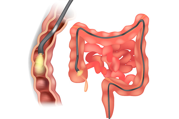
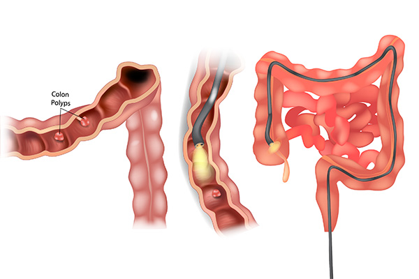
A colonoscopy involves the insertion of a lighted flexible tube, called a colonoscope, into the rectum. The tube is inserted so that the lining of the colon is visualized. Any area of the lining that appears abnormal may be biopsied; that is, a piece of tissue may be removed for analysis. In addition, growths of the colon, called polyps, may be removed (polypectomy) by the use of an electrified wire, called a snare.
A colonoscopy is generally a safe procedure but in rare cases, complications from a colonoscopy may include: a reaction to the sedative used during the exam, a tear in the colon or rectum wall (perforation), or possible bleeding from the site where a tissue sample (biopsy) was taken.

Upper Endoscopy (EGD)
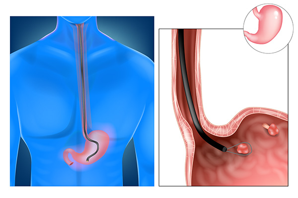
An EGD is also referred to as “upper endoscopy” or “gastroscopy”. It involves the insertion of a lighted flexible tube, called an upper endoscope, into the mouth. The tube is guided by direct vision into the esophagus, stomach, and duodenum so that the lining of the upper gastrointestinal tract is visualized. Any area of the lining that appears abnormal may be biopsied; that is, a piece of tissue may be removed for analysis. Areas that are bleeding may be cauterized to stop active bleeding or to prevent future bleeding.
An EGD is generally a safe procedure but in rare cases, complications may include a small hold in your esophagus, stomach, or small intestine. There may also be a small risk of bleeding from the site where the tissue was removed.

Sigmoidoscopy
A sigmoidoscopy is a diagnostic test performed using a thin, flexible tube called a sigmoidoscope. The procedure examines the sigmoid colon, which is the lower part of your colon or large intestine. A sigmoidoscopy may also be used to take a tissue sample or biopsy and can remove polyps or hemorrhoids. It is also used to screen for colorectal cancer.